What is Pelvic Ultrasound?
Ultrasound is safe and painless. It produces pictures of the inside of the body using sound waves. It uses a small probe called a transducer and a special gel placed directly on the skin. High-frequency sound waves travel from the probe through the gel into the body. The probe collects the sounds that bounce back. A computer uses those waves to create an image. Because images are captured in real-time (right away), they can show the structure and movement of the body’s internal organs.
A Doppler ultrasound exam may also be part of a pelvic ultrasound examination. Doppler ultrasound is a special ultrasound technique that evaluates movement of materials in the body. It allows the doctor to see and evaluate blood flow through arteries and veins in the body.
What are some common uses of the procedure?
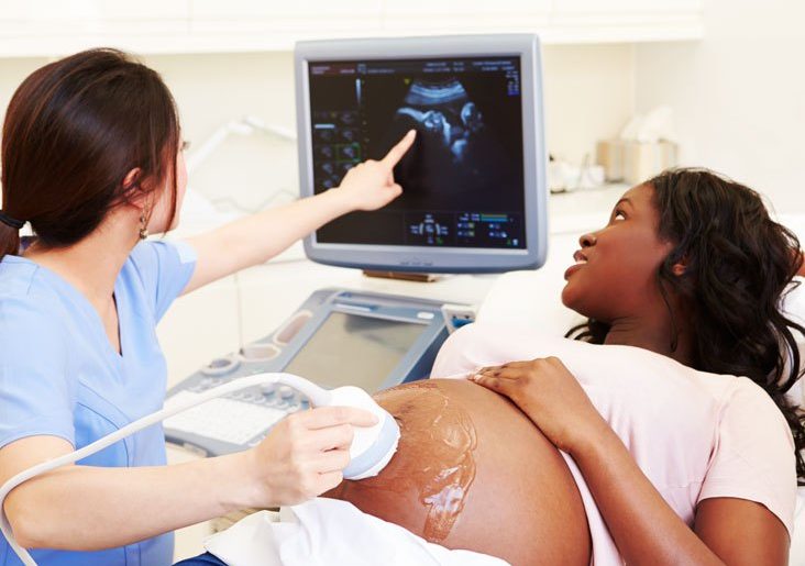
In women, a pelvic ultrasound is most often performed to evaluate the uterus, cervix, ovaries, fallopian tubes, and the bladder. Pelvic ultrasound exams are also used to monitor the health and development of an embryo or fetus during pregnancy.
Ultrasound examinations can help diagnose symptoms experienced by women such as pelvic pain, abnormal vaginal bleeding, and other menstrual problems. Ultrasound exams also help identify masses such as ovarian cysts and uterine fibroids, ovarian or uterine cancers.
A transvaginal ultrasound is usually performed to view the endometrium (the lining of the uterus) and the ovaries. Transvaginal ultrasound also evaluates the muscular walls of the uterus. Sonohysterography allows for a more in-depth investigation of the uterine cavity. These exams are typically performed to detect uterine abnormalities, uterine scars, endometrial polyps, fibroids, cancer of the ovary and the uterus.
How should I prepare?
Wear comfortable, loose-fitting clothing. You may need to remove all clothing and jewelry in the area to be examined. You may be asked to wear a gown during the procedure.
Ultrasound Procedure
A small amount of gel is applied to the area under examination and the transducer is placed there. The gel allows sound waves to travel back and forth between the transducer and the area under examination. The ultrasound image is immediately visible on a video display screen that looks like a computer monitor.
Some ultrasound procedures, such as transvaginal exams, require insertion of the transducer in the vagina. In these cases, the device is first covered with a sheath and lubricated. This examination is painless, and is often more comfortable than abdominal examination of the pelvis as a full bladder is not required.
How is the procedure performed?
For a trans-abdominal ultrasound exam, the patient will lie face-up on an exam table. Patients may be turned to either side to improve the quality of the images.
After you are positioned on the examination table, a warm water-based gel is applied to the area of the body being studied. The gel will help the transducer make secure contact with the body and enhance the ultrasound image. The transducer is placed on the body and moved back and forth over the area of interest until the desired images are captured.
There is usually no discomfort from pressure as the transducer is pressed against the area being examined. However, if scanning is performed over a tender area, you may feel pressure or minor pain from the transducer.
Once the scan is complete, the clear ultrasound gel will be wiped off your skin. The ultrasound gel does not usually stain or discolor clothing.
Transvaginal ultrasound is performed like a gynaecologic exam. It involves the insertion of the transducer into the vagina. The bladder should be empty for better visualization. The tip of the transducer is smaller than a standard speculum that is used when performing a Pap test. A protective cover is placed over the transducer, lubricated with a small amount of gel, and then inserted into the vagina. The images are obtained from different orientations to get the best views of the uterus and ovaries. Transvaginal ultrasound is usually performed with you lying on your back, possibly with your feet in stirrups as in a routine gynaecologic exam.
If a biopsy is performed, additional discomfort (due to the needle insertion) is usually minimal because the rectal wall is relatively insensitive to the pain in the region of the prostate. A biopsy will add time to the procedure.
Benefits
- Ultrasound scanning is noninvasive (no needles or injections).
- Mostly, ultrasound exams may be mildly uncomfortable, but it is not painful.
- Ultrasound is easy-to-use and less expensive than most other imaging methods.
- Ultrasound is the preferred option for the diagnosis and monitoring of pregnant women and the unborn child.
Risks
Standard diagnostic ultrasound has no known harmful effects on humans.
Larger patients are more difficult to image by ultrasound because greater amounts of body fat impair the image seen from the transducer.